Well, finally the snow has cleared and the Northeastern United States is no longer looking like the Arctic. The only snow that remains around here are the frozen remnants of the largest piles of plowed snow with bits and pieces of driveway gravel left in its wake. After a long period of hibernation, out peeks a crocus and the hints of daffodils which harken to warmer days that we strain to remember. And in a flash, people wil be complaining about the humidity and be hunting for shade! The joy of living in a seasonal climate!
As the Spring season begins, a new set of ocular symptoms appear to arise along with the emergence of the bulbs. One would not necessarily think of disease as cyclical, but we all see different ailments at different times of the year. Or maybe just more of some at different times since most diseases are not restricted by time. But before we go there, a different seasonal reminder first.
The 4th Annual Nation Service Dog Eye Exam Month
For the fourth consecutive year, Merial and the American College of Veterinary Ophthalmologists have combined to provide free eye exams for any dog officially performing a service function. Whether you are the Dalmation on the fire engine, a Guiding Eye dog or drug sniffing for Homeland Security, you are entitled to a free ocular exam by a participating ACVO diplomate during the month of May. We have participated each and every year and are happy to examine your friend once you have registered through the appropriate website provided. We have made space available on Thursday afternoons throughout the month of May for this purpose. If you and your dog fulfill the criteria, go to ACVO website (www.ACVO.org) for the link to the official registration site and then call our office to set up your free exam! Additional details are provided on the website and at the Helpful Resources tab on this website. Hope to see you soon!
Conjunctivitis
Seasonal conjunctivitis is very common around this time of year and in the fall. Most of these allergies are probably related to aerosol bourne antigens related to all the blooming plants and grasses. However, many things can create allergic responses. Most dermatologists won’t commit to an allergy diagnosis before 2 years of age in a dog, however, some reactions are seen. In addition, small lymph node-like structures called follicles will become elevated and irritated as a non-specific response to low-grade, chronic irritation. We see this response in our allergic dogs, those running through the fields like the hunting breeds, secondary to diseases like dry eye, and also in youngsters as a presumed maturation issue. All of these entities present with a history of discharge as well as redness.
If discharge is grey-white, it is usually mucus indicating irritation and inflammation. Clear tear may be present of the only type of discharge in this situation as well. If it is yellow green, this suggests bacterial infection. Brown discharge is not specific for any condition and is usually related to a pigment called porphyrin that is in the tears. This is responsible for the tear staining you may see in the corner of white-haired dogs that are tearing. Treatment therefore depends on whether the problem is inflammatory, infectious or irritative in nature. Also, local problems such as ear and teeth infections can contribute to discharge and, once treated, may resolve or diminish the discharge. The upshot….an examination is warranted to help with diagosis and drug selection.
Traumatic corneal injuries
With the weather changing, our dogs are relieving their cabin fever by racing outside and sticking their faces in bushes in search of balls, squirrels and other interesting things. This indiscriminate placement of the eye in imminent danger leads to numerous traumatic injuries that may warrant our service. Corneal lacerations, foreign bodies, simple erosions and deep melting ulcers seem to spike in the spring due to this increase in activity.
Foreign Bodies
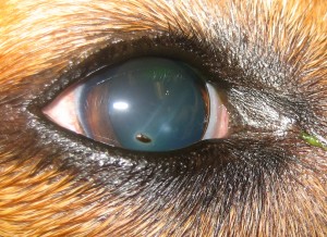
Foreign bodies can be anything that sticks to the cornea and can be a small plant seed hull to a thorn in the eye. The depth of penetration is what dictates what can be done and the prognosis after recovery. I have numerous pictures that can be unsavory of the many things that get stuck or poked into the eye. To keep things “G rated”, a small plant object is shown. This type may be flushed out or removed with a topical anesthetic. Others that may penetrate into the eye may cause the eye to leak once removed and are removed under a general anesthetic. If leakage is noted, closure or grafting could then be safely performed.

Superficial corneal foreign body
Corneal lacerations
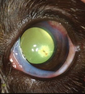
Corneal lacerations are seen most commonly in puppy versus cat altercations, however, any sharp object or tree limb can do it. These lesions are typically linear. Depending on the angle of entry, they may be straight and even or have a flap of tissue. Again, depending on the depth and angle, these may resolve with just medical treatment like a simple scratch wound or necessitate suturing if deep and/or long. If penetration into the eye has occurred, the prognosis depends on whether the intraocular structures have been involved and if bacteria have become transfered into the eye.

Corneal erosions
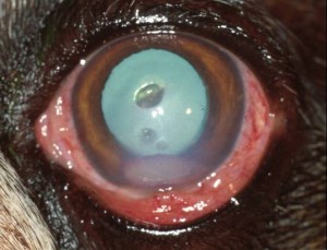
The cornea can get scratched in numerous ways but what makes the difference in healing is whether infection is present. A garden variety corneal ulceration will heal from side-to-side in a few days in the presence of prophylactic topical antibiotic, an Elizabethan collar and dilating agents if needed for discomfort. If the corneal becomes inoculated with bacteria, some nasty types will produce enzymes that digest the cornea. Depending on the severity of the bug, a hole can develop in 1-2 days that can rupture the cornea. Others may digest the tissue more slowly and the ulcer will appear more sloping than cookie-cutter in appearance. Aggressive medical and surgical therapy is required in these cases with surgery typically suggested if the depth is greater than 50% in an active ulcer. Marked discomfort and yellow-green discharge are usually noted in these cases. We may talk about surgical treatment and the postoperative appearance later, but here is an example of a typical “melting” corneal ulcer.

Deep ulcer with inflammation
All for now….see you soon, and don’t forget to get your Service Dog examined!


