Tired of the rain yet? The grass is soaked, basements are flooded, rivers are swollen. Where is the summer sun? And the humidity hasn’t been much fun either. With the non-stop rain here in Connecticut, everything seems to be dripping. That includes lots of our canine patients as they run in from the parking lot! We also see lots of “weepy” eyes where the complaint is primarily a clear, sometimes colored, discharge. Let’s take a look to see what may be behind the scenes with this presentation.

When our patients present with clear discharge, my first question is whether we are making too much tear in response to irritation or whether the outflow pathway, or tear ducts, are compromised. One could presume that in the former instance discomfort would be a feature. This will be manifest as squinting, pawing, rubbing and/or redness to the eye or around the eye. In the latter, discomfort may not be a big feature even with lots of moisture pouring out of the eye.
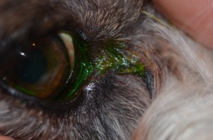
Diseases with discomfort that present with tearing are many as this is a non-specific sign. However, the nature of the discharge would suggest that infection is not playing a big role otherwise we would see yellow-green discharge. Similarly, significant irritation with marked inflammation my have more mucus or a grey-white discharge. For example, the hallmark of conjunctivitis is discharge but usually one with color. Irritation from foreign bodies or offending hairs may create corneal erosions that are irritating but not infected and thus a clear discharge along with discomfort will be the result. Issues where the lid conformation is abnormal create surface irritation due to the adjacent fur rubbing on the eye. Tearing can then occur during this process which can spill onto the face either directly from the irritation or from a wicking effecting where the tear spills over the lid margin onto the side of the face adjacent to where the lid rolls in. Entropion is the technical name for lids that roll in as a result of genetics (primary entropion) or secondary to globe retraction from surface irritation like a corneal ulcer (spastic entropion). Resolution of the primary entropion by surgery to roll out the lids or appropriate medical or surgery treatment to cure the irritant that causes the spastic entropion will stop the tearing. It’s all connected! Typically a good ophthalmologic exam will reveal the problem.
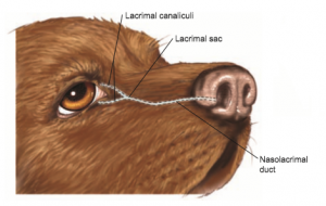
If upon inspection and testing we rule out surface disease then investigating the outflow system is warranted. Tears are produced by glands that exit through the conjunctiva onto the surface of the eye. These tears then need to be “removed” either by evaporative drying or out through the ducts that are located in the inner aspect of the lids and pass under the surface of the conjunctiva and skin, shortly through bone and into the nose. Most of our patients, except the rabbit, have an upper and lower duct that then join together before exiting the nose. The opening to the duct is called the puncta. Obstruction or narrowing anywhere along this pathway may make the path of least resistance to go over the lid margin and onto the face. This is commonly seen at the inner aspect of the eyelids rather than the outer aspect by the ear. The length of this duct varies with species and breed and thus different issues can affect its passage depending on the type of animal.


The lower puncta is easily viewed here as the small non-pigmented circle just inside the pigmented lid margin.
Two tests are used in the exam room to evaluate the patency, or intact nature, of the duct system. The first is a passive test, called a Jones Test, where fluorescein dye is placed on the eyeball to see if this dye flows into the nostril region. This dye glows with a blue light and can be easily seen. This test is easy to perform and will tell you if the “plumbing” is working. However, a negative tests does not necessarily tell you that the duct is not patent since flow rate can be slow or the duct may be narrowed which affects the time it takes for the dye to get to the nose. Also, it does not tell if the flow is going through both ducts near the globe initially. Another benefit of the test is that the dye may wick onto the face quickly in some dogs with hair issues that draw the tears onto the face before they get into the puncta.
The second test is more active called nasolacrimal flushing. In this test, a small cannula or catheter connected to a syringe of flush is placed in the upper or lower puncta and gently pressed. The fluid should come out the other puncta and, when obstructed with a finger, out the nose. This should occur with minimal resistance. Most dogs and some cats are amenable to this with just a topical anesthetic. We get information as to whether the openings are intact, if flow out the nose is present, if there is any resistance to outflow and potentially release of a loose blockage.

Fluorescein dye wicking onto the face instead of going into the puncta due to hair and lid anatomy.
A common entity we see in young dogs is an abnormal development of the lower opening or puncta. An imperforate puncta is when a sheet of tissue is present over the opening due to improper formation at birth. The duct is typically present just under the surface. This is much more common to be seen with the lower puncta. Opening this puncta under a short anesthetic event can improve and sometimes resolve the overflow of tear in the affected eye.
Cats rarely get imperforate puncta but commonly get herpes infections. One manifestation of herpes when acquired as a neonate is the development of adhesions of the conjunctiva to the cornea, third eyelid or itself called symblepharon. Tearing can be noted if these adhesions are associated with the puncta or tear ducts even if the infection is cleared or inactive. Basically these adhesions are scars from the infection. Surgical resolution is typically unrewarding since they recur, however, this is usually good news if all we are left with is tearing. A full discussion of herpes is not necessary here.
Obstruction downsteam of the puncta is less common. Dental or sinus disease can theoretically present with tearing due to the proximity of these structures and the tear duct as it passes into the nose. Mucus plugs are occasionally flushed out but are not too common. Other symptoms may be present if there is trouble downstream of the puncta.
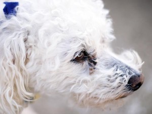
A result of the tearing that bothers many owners, but rarely the dog or cat, is the brownish-red staining of the fur in the wet region as seen in the first photo above. A pigment called porphyrin in the tears reacts with the fur and creates a brown tinge that is very obvious on our white dogs such as Poodles, Bichons and Maltese. This discoloration can happen at any time and is not indicative of any disease process. There is no specific product, diet or medication that resolves this issue much to the chagrin of many. The good news is that this is a cosmetic issue rather than a medical one. The degree of staining may decrease if there is a primary issue when can address that will minimize the overflow.
That’s a wash here! Maybe by the time you have read this the sun will be out!


