A common comment veterinarians hear is about how our job is so difficult with a patient that cannot tell us what is wrong. Well, sometimes less is more! Our patients talk to us in different ways with clinical signs and symptoms that help us determine what and where the problem is without the confusion of speculation and interpretation and emotional embellishment of those features that we all do as humans. We use our senses and powers of observation along with listening to the heart and lungs and palpation of the body to get most of the answers in general practice. Blood screening, imaging and other minimally invasive tests are then usually performed to see if we can support our observations with supportive data from these tests.
In many ways, what we do as ophthalmologists is easier than in other disciplines because most of the information is literally right before our very eyes. We can see the cornea and lens to assess its relative health and clarity. The eye is the only structure in the body where you can look directly at a major blood supply to see if the tissue is being supplied with blood to keep it healthy (in the retina). Everything from the movement of the eye to the responses to light can all be directly evaluated. Interpretation of dye staining and measurement of eye pressure, for example, are usually to support a suspicion of something we can see clinically. The trick is that the eye is very small, requires a steady hand and an unwavering gaze to be evaluated. Throw in a mobile patient that won’t respond to “Look up!” and you have the challenge of being a veterinary ophthalmologist.
Surgery may be a little easier since the patient is finally immobile for our examination, however, manipulation of needles and suture that are crazy small is a special skill all its own. And a scar in the skin is no big deal but the cornea needs to remain as clear as possible for it to do its job. So surgical trauma has to be minimal in cases of corneal or lens disease to keep them or other tissues functioning at their best after treatment. How do we do all this? On top of years of study and inherent or developed skills to keep that hand steady, we are reliant upon some pretty sophisticated equipment to do our job. So here is a brief rundown of some of the stuff we use to help your critters out in the world of veterinary ophthalmology.
The equipment used in human and veterinary ophthalmology is really quite interesting. We need high intensity light sources with focused beams and crystal clear magnification to be able to evaluate and treat all of the diseases that present themselves. The two workhorses of the exam room are the slit lamp biomicroscope and the indirect ophthalmoscope.
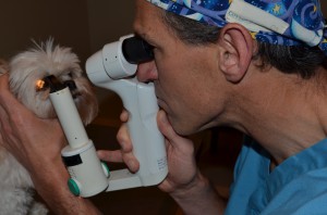
The slit lamp biomicroscope in use
The slit lamp is an instrument used to see the front structures of the eye like the lids, cornea and the lens. Ours has three different levels of light intensity and two levels of high magnification. In addition, the beam of light comes in four shapes, a circle and 3 vertically aligned beams of light that are progressively thinner in width from 0.8mm to 0.1mm. In addition, we can use a cobalt blue filter on the light source to help with observing fluorescein dye on the cornea to highlight ulcers. Along with good lighting and magnification, the benefit of this instrument is that we can swing the light to the side while looking at the structure from straight on. With a slit beam of light, we can see a cross section of a clear structure that then allows us to examine the depth or placement of a lesion. For example, if there was a mark on your glass door, you may look from the side to see if the spot was on the outside or the inside before you started cleaning. With a pane of glass 1/4 inch or greater thick, you could easily see the dirt was on the outside plane and not the inside and thus walk outside to clean it. The same process is applied when looking at the cornea only it is 0.5mm thick! So if we want to see how deep a thorn has punctured into the eye before we pull it out or see how deep a ulcer is to determine if it is a medical or surgical condition, the slit lamp is your instrument.
The slit lamp is also used to observe the size and location of cataractous opacities in the lens or the severity of inflammation inside the eye. With intraocular inflammation, called uveitis, the beam of light is used to see if we can see the light cut through the fluid inside the eye and create a stream of light like a flashlight on a foggy night. A normal eye should not have a stream of light, an inflamed one does and can be graded to assess severity. Our handheld version is similar to the large instrument you place your chin into at the human ophthalmogist to assess these same features. Our dogs and cats just can’t get their noses and chins to sit just right in this machines! Our version can be used by doctors visiting out-patients or doing rounds in a hospital situation where a cumbersome large machine or a non-mobile patient does not allow its use.
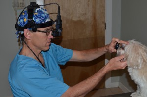
Viewing the retina with an indirect ophthalmoscope
The indirect ophthalmoscope is typically used to evaluate the back of the eye, primarily the retina. By using a focal light source and a second lens to focus the light, we can screen the entire retina with a single gaze. Different powered lenses can be used to magnify areas of interest or increase the field of view depending on what you may be looking for. These lenses can help even with small pupils so we don’t necessarily have to dilate the pupil if we don’t need to see in the far periphery. This exam can be a bit uncomfortable for you and I but most dogs and cats sit just fine for us to look.
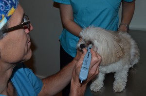
Applanation tonometry in the dog
We do numerous tests in the exam room to evaluate everything from tear production, tear quality, tear duct function, corneal intergrity, detection of bacterial infection or inflammation and eyeball pressure assessment. Glaucoma is a very common disease and intracocular pressure measurement is performed regularly to determine this value. High values equal glaucoma, low values are suggestive of inflammation with a wide range of normal in between that varies slightly with species. There are also many different tools used to measure intraocular pressure which is called tonometry. We perform what is called applanation tonometry. In this test, a finely calibrated instrument with a sensitive mobile tip is tapped upon the surface of the clear cornea. The tip will depress slightly and the amount of pressure needed to flatten the cornea is determined by a microchip in the device to produce a number that equates to how hard or soft the eyeball is inflated. When pressure is high inside the eye it creates all kinds of problems that lead to vision loss. This is a very important test that is performed routinely on most exams.

Phaco machine and handpiece
Cataract surgery is a commonly performed procedure that is done on an elective basis to return vision to your friend if the lens has become opaque. This is performed by a technique called phacoemusification where high-frequency ultrasound power is used to break the lens up into small pieces while concurrently keeping the eye inflated with liquid and sucking out the particulate material that is created as the lens disintegrates. Our machine, an Alcon Legacy 20000, does a wonderful job of that and you can see in the picture the handpiece that is placed inside the eye with the computer screen that displays all our settings that affect power, vacuum, suction rate, etc. It really is a fascinating surgery to perform and watch. A video in our Helpful Resources page and a prior post about cataracts in the dog have additional details and images about this disease and procedure. The Legacy (we call her Alice!) is our workhorse for this procedure and we take great pride in keeping her in tip-top shape for this wonderful procedure.
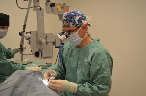
Using the operating microscope
To perform most of our intricate corneal and lens surgeries, be it cataract extraction or repair of deep corneal ulcers, we need magnification to allow visualization of the tissues. Our operating microscope is a beautiful piece of equipment with a large arm that swings the microscope over the surgical table and directs the beam into the eye. We can zoom in and out, move up and down or from side to side with a foot pedal to keep our hands free and sterile. The intensity, width and focus of the beam can be adjusted to maximize our ability to see at any depth and degree of magnification. This expensive tool is mandatory for doing these surgeries not only to see the structures but also the fine suture we use. Sometimes it looks like there is nothing in our hands from the casual observer that is not looking through the scope as well. That’s why I frequently have an assistant scrub in with me to watch though the assistant’s eyepieces and pass instruments to me during the procedures.
These are only some of the instruments and technologies that are used in ophthalmology to allow us to see what your pet sees. Boarded ophthalmologists are trained to use these tools, understand their nuances and limitations, and then apply the knowledge they have obtained to aid in diagnosis and treatment of your pet’s ocular disease. As the saying goes, it’s not just the wand but the magician behind it as well! But it sure helps to have some nice wands!


